Wednesday 12 February 2014

Science journalism needs to change - A rant

The way science is reported in mainstream media needs to change. There are some excellent science reporters in the field and I am not denying that, but by and large much of the science that ends up as headlines resembles celebrity gossip columns with attention grabbing headlines, usually containing the word breakthrough, that over-arch on a small bit of evidence. This, to me, is a double edged sword. Sure, it raises the profile of science and scientists but at the same time it's becoming a 'boy who cried wolf' scenario with the general public seeing a breakthrough every other day. I cannot help but feel this will erode the publics trust in either the reporting of science or science itself, either way science loses. Look at this article from the BBC for example running the headline "Alzheimer's breakthrough hailed as 'turning point'". Upon a further inspection of the article, and indeed the original journal article itself, we see that researchers have given mice with prion disease (a disease which bears some similarities to Alzheimer's disease but is distinct from it) a "drug like compound" and have seen symptoms alleviated. So they gave mice with a disease that isn't Alzheimer's disease a drug-like compound. Call me a sceptic but this has a very very long way to go before we can get excited about this treating people with Alzheimer's disease

To begin with the disease they tested isn't even Alzheimer's disease, it's a related disease in the class of 'proteinopathies'. Second, it is in mice, Alzheimer's disease has a very poor track record when it comes to seeing benefits in mice translate into the human disease (see the recent clinical trials here  and here). So ultimately, I am of the opinion that the terms "breakthrough" and "turning-point" and entirely unwarranted here. The article could have run with a title such as "New drug-like compound alleviates symptoms in a mouse model of Prion disease" which sure, doesn't have the emotive response of words like "breakthrough" but is accurate and not over-arching. Isn't that what reporting is meant to be? On that note, titles/articles should not only mention the limitations I have just mentioned but also the side-effects. The BBC article mentions them briefly with this "The compound also acted on the pancreas, meaning the mice developed a mild form of diabetes and lost weight", upon reading the paper we see that the experiment had to be cut short and the animals killed because their weight loss was so unhealthy it was deemed unethical to continue the trial. Again, call me a sceptic, but this seems like it would limit the applicability in human trials.

It may seem like I am picking on one article here but unfortunately articles similar to this are the rule, rather than the exception. Science needs to be communicated to the public. Much of the time the public funds it and it usually affects them, but reporting needs to be done accurately without the over-inflated life changing benefits. In the paper above, I personally would have a section explaining how difficult and expensive it is to take a finding such as that one and investigate the potential to treat the human disease and call for financial support for the research. 

What is the solution to all this you ask? In the long term, the business model of mainstream reporting needs to change, but in the short term, blogs, science websites and the like are doing an excellent job of providing multiple points of view on a particular paper. "Groundbreaking" articles like the one above attract a fair bit of attention from other scientists and so a quick Google search will no doubt bring up some alternative opinions which are worth checking out. There are websites such as Sciengage (for which I blog) that are attempting to bridge that gap and are doing a great job of it. In the short term, if you read a "breakthrough" that really interests you, I would encourage a quick 5-10 min Google search to see if others have written about the same article or take to social media, find a scientist, and ask them if the conclusions are warranted (that is precisely how I came across the above article, someone asked me on Twitter).

Thursday 5 September 2013

Mind control

As you may or may not have noticed, my blog posts have been rather lacking recently. I whole-heartedly blame my pesky PhD getting in the way of my blogging. Never fear. I shall resume regular postings after a conference this week. In the meantime, here is a very quick note about a pretty cool finding that came out recently.

This has not been published as a paper yet but the researchers did a press release and my write up will be based on their press-release (which can be read here).

Two subjects were involved. The first (subject A), had a complex EEG cap on his head (this measures the electrical activity at many places throughout the brain by attaching very sensitive sensors to the persons head). The second subject (subject B), on the other side of campus, wore a piece of equipment that performed "transcranial magnetic stimulation", which, as the name suggests uses magnetic fields to stimulate an area of the brain through the skull. This piece of equipment was placed directly over the part of the brain that is known to be responsible for moving the right hand.

Subject A sat in front of a screen with a simple computer game on it but had no controls. The game required pressing "fire" at the right time to shoot something. Subject A was told to, not move his hand but imagine pressing his right hand to press the fire button (but of course had no controls to press). The brain activity of subject A was recorded by the EEG machine and was encoded and transmitted, via the internet, to the equipment on subject B's head. Once there, the equipment on subject B's head mimiced the exact brain activity that occured in subject A's brain.

Schematic of experiment set-up taken from here


The end result?

When subject A imagined pressing fire, subject B involuntarily pressed fire without any control of his hand. In other words, subject A's thoughts were being used to control subjects B's body movement. Demonstrating brain-to-brain control, across the Internet!

This is the first ever human, non-invasive, brain-to-brain interface.

Science.
Is.
Rad.

Fact.

Here is a (albeit rather dull) youtube video put out by the researchers.


This researchers a quick to reassure the public that this is very far away from controlling someone's mind against their will.

claimtoken-52310c2f859e2

Friday 19 July 2013

Levitating objects .... with sound!

It has been known for 100 years that sound waves can exert a force (the acoustic radiation pressure effect) but not until now has this principle been used to manipulate physical objects.

I'll keep this post pretty short because the maths involved is pretty obscene but for those who are slightly masochistic and would like to take a look, the paper by Foresti et al. was published yesterday in PNAS and can be viewed here.

The basic principle used here is there is an emitting platform (that as the name suggests emits the sound wave) and then placed opposite that, there is a reflecting platform and these platforms are placed at particular places based on some complex math. The figure below is a diagram of the apparatus taken directly from the paper.

Setup of droplet manipulator (Foresti et al. (2013)
I won't get into it but take my word that the design of this system and the calculations involved is an example of some beautifully thought out elegant science and props to those involved in its development.

Now, it's all very well to make an object levitate using a sound wave, but they go one step further and actually move the object with these sound waves which again has some pretty complex equations behind it. The sound emitting mechanism is made with piezoelectric crystals, which shrink or stretch depending on the voltage applied to them and hence can alter the sound wave produced quite finely and accurately.

As a proof of principle they did several awesome things! First, I will show you the video of them moving and mixing droplets of water seen in the video below:


Then as true scientists, they wanted to make this useful for something very important to most scientists ... Making coffee ... Check it out:


Yep, they are mixing a granule of instant coffee and water in thin-air to make a drop of coffee! That video is slowed down by 100-times so you can see the fine details.

Next they took a step larger:


I don't know about you, but I find that pretty incredible!

The applications for this are not as wide-ranging as you might think unfortunately. The object being levitated and the sound waves have a very intimate relationship, the diameter of the object needs to half of the wavelength of the soundwave, so big objects are out of the question at this stage. However, it does have some exciting applications for chemistry as many chemical reactions and science experiments are influenced by the chemistry of being on the surface of something so we can now investigate questions free from surface interference.

So aside from making coffee in mid-air, the benefits of this amazing technology remain to be reaped by scientists ... For now.


Foresti D., Nabavi M., Klingauf M., Ferrari A. & Poulikakos D. (2013). Acoustophoretic contactless transport and handling of matter in air, Proceedings of the National Academy of Sciences, 110 (31) 12549-12554. DOI:

Friday 5 July 2013

How to grow a liver in a petri dish and save lives

Liver donors are extremely rare - In 2011, 5,805 liver transplants were completed in the US and that same year, 2,938 people died waiting for one. Needless to say, an area that needs attention. In a first of a kind study by Takebe et al. published in Nature this week we may be a step closer to solving this problem.

The group has used human stem cells (I'll go into how towards the end) to create what they have dubbed 'liver buds', that is basically miniature human livers (about 4mm in diameter). They have then taken these liver buds and surgically implanted them into mice. The surgically implanted buds were able to substantially prolong the life of mice with liver failure! This is itself is a pretty amazing feat of science! Taking some human cells, growing a mini liver, throwing into an animal with liver failure and they live longer!

The liver buds not only prolonged the lives of these mice but incorporated themselves with the mouses blood system and continued to grow after the implantation. The buds took on many features of a fully-functional liver such as producing and sending out liver-specific proteins and signals. The figure below shows the implanted bud (in the dotted area) gaining blood vessels over time (shown by the increase in red in the dotted area).

Figure showing vascularization of liver buds (reproduced from Takebe et al. (2013))
These mini livers were the result of combining 3 types of human stem cell. First, pluripotent stem cells were converted into cells that are programmed to express liver specific genes (this is done by given the cells signals that trick them into thinking they're growing in a liver). Next, they combined these liver-specific cells with some endothelial cells (the kind of cells that make your blood vessels) and some mesenchymal stem cells (these can make bone, cartilage and fat). Over time, these groups of cells interacted and self-organized into the liver buds seen above. Below is an illustration of the process of creating the liver buds (panel A) but also a visualization of them forming over time (panel B). You can see how they start as a large smear and as time progresses they come together in an organized fashion.

Figure showing A) An illustration of the methodology of the experiment and B) showing the formation of the liver buds over time. (Reproduced from Takebe et al. (2013))
The figure below is what is called a "survival curve" which illustrates the rate of death of the mice in different conditions. Every vertical drop in the line is a mouse (or mice) dying. So a flat line means the group is surviving, a rapidly decreasing line shows they a dying.

There are only two lines you need to worry about. The dotted line at the very bottom is the group of mice that had the procedure done but didn't have a bud implanted (the control group), and the solid black line at the top is the group of mice that had the procedure done and did have the liver bud implanted.

Survival curve (reproduced from Takebe et al. (2013))
This shows that when the mice don't have the treatment, over a 30-day period, they die fairly rapidly. However, when they receive the bud implants, over the 30-day period, only ONE mouse died!

I don't know about you but I find that pretty amazing and VERY exciting. Of course as with most first of a kind studies, it needs to be replicated and further studies done but the implications this has for not only those with liver damage, but with any organ damage is huge and every exciting.

PS. For those of you who do not belong to an institution and cannot view the original article there is a summary here.

Friday 28 June 2013

A boy and his atom ...

Today is going to be a very short post, but not much about a new, cutting edge piece of science! More using the incredible technology science has to offer... to create art!

This was a video created by IBM and is officially the worlds smallest stop motion film. It was created to celebrate IBM creating the smallest magnetic memory bit (comprised of only 12 atoms, the current norm is around 1,000,000 atoms, so not bad work!). So to celebrate this, they used the technology in their lab to create a stop motion film .... of individual atoms!

The project was "filmed" using what's called a scanning tunnelling microscope (which was actually invented by IBM researchers who got the Nobel Prize in Physics for it in 1986). Now, your normal everyday microscope won't cut it to see atoms, in fact you can't even use light to see atoms, so the scanning tunnelling microscope works by gently moving a super-fine needle above atoms of the material being "imaged". As the needle moves over a particular atom, a mini-chemical bond is formed between the atom and the needle tip, this can be measured through the needle. The needle tip is moved ("scanned") from left to right, right to left, left to right moving down the material. When something is higher (closer to the needle), then a stronger bond can be detected, but if the atom is far away from the needle then only a weak bond can be detected. As the needle "scans" the material to be imaged, all this data is then compiled into a viewable image!

For this film, the researchers used the bond formed between the tip and the atom to actually move some atoms around, to create a picture! Then they moved them again and took another, and so on to create this amazing stop motion film.

Each small globe is ONE INDIVIDUAL ATOM of carbon monoxide. Think about that for a second!


IBM did a (fairly) easy to follow video on how they made the atoms move, they may do a better job than I did:


And that's it! Hope you enjoyed it, fairly simple concept but done really really well in my opinion!

PS. Apologies to all physicists reading this, hate mail can be addressed to me personally :)

Friday 21 June 2013

A viral infection of the eye that can cure blindness?

A very controversial area in science is gene therapy with the use of viruses (it really shouldn't be that controversial thou). I realise it sounds quite scary to be injected with an actual virus, but gene therapy using a virus as a tool is a remarkably elegant (and relatively safe) technique which deserves some time to clarify. A search for 'gene therapy' in Pubmed gives back just shy of 40,000 peer-reviewed scientific papers, so it is clearly an exciting, well-researched area of science.

Now, a bit of background. Viruses are sneaky little buggers and have evolved over time to sneak past your normal immune defences and infiltrate your cells, and from there trick the cell into making more of the virus. Normally this is a bad thing such is the case with HIV or the more common virus, the flu. So what happens, if we take away all the parts that allow a virus to make us sick and give it some specific DNA that a patient might be missing. Then the virus will go, sneak into the persons cells and trick the cell into making proteins from that DNA, but the proteins will be good for the patient and replace an otherwise missing protein.

There are many diseases that are caused by a mutation in the DNA which then means the protein isn't made in the patient, so if we could somehow deliver the protein (or at least the blueprints for the protein, DNA) into the cells then we could replace what was missing.

Gene therapy using an AAV virus. (Source: Wikipedia)

Gene therapy has been successfully and safely used to treat many diseases such as chylomicronemia, chronic lymphocytic leukemia, and multiple myeloma and a clever technique has recently been published to advance the use of this technique to treat certain types of blindness.

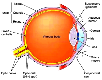
Inherited forms of retinal diseases afflict 1 in 3000 people worldwide and are primarily the result of mutations encoding proteins in the retina of the eye. Current gene therapy treatment for these diseases involves a needle piercing the eye and penetrating the retina itself to successfully deliver the virus. The diagram below shows just how far back that needle would have to go. The process requires hospitalization, general anaesthetic and runs a real risk of damaging the retina itself. This is because the special viruses usually used in gene therapy (called AAV) cannot get that far back into the eye normally and so need to be injected directly there.


Source: http://encyclopedia.lubopitko-bg.com

A group from Berkeley University has made use of the high rate of evolution in viruses (because they reproduce so quickly) and used directed evolution to "evolve" a virus that has the capabilities to get that far back in the eye. To do this, they took the standard AAV virus and injected it into the gel layer at the outer eye (the vitreous body in the diagram above), they then waited a while (a week) and collected the cells in the retina of the eye to see if any viruses made it that far back. There were some! So they let these few replicate into huge numbers again and repeated the process and repeated it and repeated it, each time only continuing with the viruses that somehow had the ability to reach the back of the eye, therefore selecting the viruses with the desired ability. At the end of all this they found 48 AAV viruses made into the back of the retina and 2 thirds of them were the same virus, they dubbed it 7m8.

To test if this virus could then be used to treat a disease, they used two mouse models of retinal degeneration caused by malfunctioning proteins in the retina and split each disease model into two groups. In one group they injected the virus, packaged with the DNA required to make the missing/malfunctioning protein, and the other group received the virus by itself without any extra DNA packaged into it. The mice with the 7m8 loaded with DNA had great vision improvements and a protein analysis proved that the retina was in fact producing the previously missing protein.

Immunostaining for RS1 protein (Dalkara et al. 2013)
The above figure is looking for the protein that is normally missing in the one of the diseases (RS1). Panel A shows a picture of the diseased retina and there is no RS1 protein there (you can tell from no greenness), panel C shows what a normal non-diseased retina should look like (lots of green) and in the middle (panel B) we have a diseased retina that had the injection of the virus with the DNA and as you can see it looks just the normal retina in panel C! Magic! Lots of green!

Now you might be thinking? So what? They still need an injection in the eye to get the virus in the first place, and you'd be right! However, injecting into the vitreous gel layer is a simple procedure, done at your local doctors practice, under local anaesthetic and runs very little risk of damaging the retina and this makes it a much more practical solution to those rendered blind from these diseases. Furthermore, this study, as a general concept, paves the way for developing more AAV viruses that are specialised to reach certain tissue or organs.

The study was published on the 12th June in Science Translational Medicine and can be viewed here.

Friday 14 June 2013

The power of chocolate (and electrostimulation combined with physical therapy)

It will be a fairly short post this week as my PhD has kept me fairly busy with performing brain surgery on mice so spare time has been few and far between.

Building on last weeks post, that looked at the ability to control machinery (such as prosthetics) with the mind only, which has exciting implications for physically disabled people. This weeks paper again has implications for physically disabled people (primarily spinal injuries).

Around half of all spinal injuries result in chronic paralysis in humans and as such there is considerable research into the area to give the patients their movement back. The paralysis results from the injury to the spine actually severing the nerves that connect a particular area of the body with brain, so that they can no longer communicate (hence they loss of movement but also feeling in some cases also). Advances in this area would change many lives.

In a landmark paper published in Science last year (here) a group in Zurich has made a giant step forward in regaining locomotion in spinal injuries. The study was done on rats and used a recovery regime that combined physical therapy, electro-stimulation and injections of a special combination of chemicals... oh, and chocolate.

The rats spines were almost completely severed (a small amount of tissue remained intact but all nerves were severed and they had no control of their hind limbs, seen below). Then the group of rats were split into two groups, one received there aforementioned therapeutic regimen and the other did not (acting as a control).

A video showing the rats inability to walk after the spinal surgery (van den Brand et al. (2012)).

Disclaimer: Now, the for the scientists reading this, the following explanation will butcher the incredibly elegant science involved but it is a necessary evil.

The rats that were receiving the treatment initially were "encouraged" to walk by initiating the reflex walking response. This was done by placing the rats in a specially designed harness (pictured below) and placing them on a treadmill. When a foot begins to drag on the treadmill it stimulates a reflex movement that is akin to taking a step but doesn't require the nerves in the spinal chord be 100% intact. This was the first physical therapy aspect (called "treadmill trained). Another aspect of physical therapy used the same harness and placed the rats on a runway type arrangement (the platform pictured below) and the harness was used to hold them up but provided no directional movement assistance. The second paradigm looked for voluntary movement of the legs rather than the reflexive movements elicited with the treadmill. The second form of physical therapy was called "overground" training.

Source: EPFL (via National Geographic)
The regime also included electric stimulation over the area where the nerves were severed which should act to encourage new nerve growth/connection over the area in question and the same area also received injections of a tailored cocktail of chemicals (for the nerds out there it was serotonin and dopamine agonists, 5-HT1A/7, 5-HT2A/C and D1 respectively) which served to assist in the encouragement of neuronal connections and comunication.

Figure taken from van den Brand et al. (2012)
The above figure essentially shows active nerves in the spine around the area of injury. The key things to look at are first, the marked reduction between intact (no injury) and non-trained (a control), and then look at the huge gains from the overground trained group. Not quite like a normal rat (intact) but a whole lot better than non-trained. You can watch a video of this in the final at the bottom of this post. This graph also shows that treadmill training alone is not enough, the overground training needs to be included to get the full therapeutic effect.

The end result? The rats regained control of their legs! In the final phase of the study the researchers placed chocolate at the end of the stair-case (in the overgound training) and noted that the rats were near on SPRINTING to get the chocolate (albeit still in their harness). After this complex course of therapy the rats regained full voluntary movement of their hind limbs but struggled to walk on fours completely unaided indicating there is more work to be done.

The researchers did a video explaining some of the concepts which I have included below.


Once again ... Science is rad! Till next week

- Si